Non-invasive, precise, and constantly evolving, ultrasound technology has revolutionized modern clinical diagnosis. Its use is now essential in numerous medical specialties, allowing for highly detailed three-dimensional images of the human body and other animal species.
The origins and the first attempts

The first theoretical approaches to ultrasound date back to the 18th century, when the Italian physicist and biologist Lazzaro Spallanzani studied the behavior of bats. He observed that these animals were able to fly and orient themselves perfectly even in total darkness.
Through a series of experiments, he deduced that bats navigated using their hearing, and he hypothesized a spatial perception mechanism based on sound echoes, known today as echolocation. This principle represented the starting point for other scholars who, centuries later, would apply it to the development of more complex technologies such as ultrasound and sonar.
One hundred years later, in 1880, Pierre and Jacques Curie analyzed a revolutionary physical phenomenon: the piezoelectric effect. They discovered that certain crystals, such as quartz, could generate an electrical charge when mechanical pressure was applied to them. Conversely, when an electric current was applied, these crystals vibrated, producing mechanical waves, including high-frequency sound waves.
This discovery led to the generation and production of ultrasound, a fundamental element in the operation of ultrasound transducers. Even today, the piezoelectric effect is used in modern ultrasound to capture acoustic echoes and transform them into diagnostic images.
Modern developments
In 1940, American physicist Floyd Firestone devised the Supersonic Reflectoscope, the first ultrasound device for echo imaging, initially intended to detect internal defects in metal parts.
The first attempts to apply ultrasound in medicine occurred in the following years, initially for therapeutic purposes (such as diathermy). Between 1941 and 1942, the Austrian neurologist Karl Theo Dussik, in collaboration with his physicist brother Friedrich, was one of the first researchers to obtain ultrasound images of the human body, delineating the cerebral ventricles.
Also in the late 1940s, physician George Ludwig first applied ultrasonic energy to the human body at the Naval Medical Research Institute in Bethesda , Maryland. During the same period, British-born physicist John Wild used ultrasound to assess the thickness of intestinal tissue.
The change in the 1950s and 1960s
In the early 1950s, ultrasound began to be increasingly used in hospitals, thanks in part to the introduction of A-mode (Amplitude Mode). This technique allows the visualization of ultrasonic echoes as vertical peaks on a horizontal graph, where the peak height represents the echo’s intensity, position, and the depth of the structure. Although it provided one-dimensional information, A-mode was fundamental for locating structures and measuring distance.
In the 1960s, B-mode ( Brightness Mode ), or two-dimensional imaging, emerged, where each echo is visualized as a point whose brightness is proportional to the echo’s intensity. Sequential scanning of the transducer allowed for obtaining cross-sectional images of the body. The work of Ian Donald and Tom Brown in Scotland, with the first clinical prototypes, made it possible to visualize abdominal masses and improve obstetric diagnosis, opening new opportunities in medicine.
In the 1970s, the first real-time devices were introduced, enabling dynamic imaging and making ultrasound faster and more interactive. This feature contributed to the widespread acceptance of ultrasound as a first-line imaging method.
In Italy, for example, there was a +350% increase in the use of ultrasound between 1975 and 1985, especially in gynecology and internal medicine.
The arrival of Doppler and color Doppler
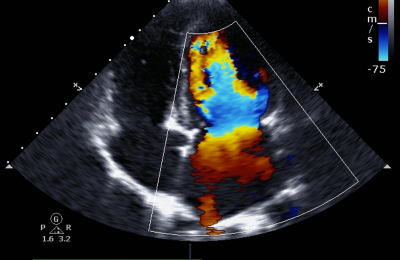
From the 1980s onwards, ultrasound also extended to the abdominal, cardiac (with echocardiography), musculoskeletal and vascular fields, thanks to improvements in image quality and the possibility of measuring blood flows with Doppler.
Pulsed-wave Doppler allows for real-time analysis of blood flow. The next innovation was color Doppler, which overlays color information onto the two-dimensional grayscale image, encoding the direction and average velocity of the flow. This development has been fundamental for the diagnosis of arterial stenosis, venous thrombosis, valvular insufficiency, and also in obstetrics, for the study of fetoplacental circulation.
The global Doppler ultrasound market was valued at $3.35 billion in 2023 and is projected to reach $5 billion by 2032, with a compound annual growth rate (CAGR) of 4.56%.
Three-dimensional and 4D ultrasound: new diagnostic frontiers
Over the past twenty years, 3D and 4D technologies have been introduced that have radically changed the visualization of anatomical structures. 3D ultrasound allows for the reconstruction of a volumetric image from two-dimensional sections, while 4D adds the temporal dimension, showing the movements of the fetus or the structures being examined in real time.
Today, over 60% of advanced gynecological centers in Europe use 3D/4D systems for fetal morphology assessment and early diagnosis of anomalies. Hospitals are the primary users of this equipment, representing approximately 50% of the market.
Portable and point-of-care ultrasound: the new era of immediate diagnosis
The development of portable ultrasound and devices connected to smartphones or tablets has made it possible to use bedside ultrasound, that is, even outside the hospital environment.
Read Also: The handheld ultrasound scanner: the revolution in immediate diagnosis
This revolution has a significant clinical impact, especially in emergency medicine, intensive care, and general practice, where point-of-care ultrasound (POCUS) enables faster diagnoses. According to a study published in Critical Care Medicine (2020), the use of POCUS reduced diagnostic times in critically ill patients by 40% compared to the traditional approach.

I am a final-year MBBS candidate. After years of watching patients misunderstand their own diagnoses, I started writing — because accurate health information should not require a medical degree to understand. I specialise in health, health tech, and public health content, combining clinical training with a passion for making medicine accessible to everyone.”